Cerebral aneurysms
This page explains in simple terms what a cerebral aneurysm is and what happens when one ruptures. It accompanies my own story. It is based on the resources recommended by the Neuro Patient Resource Centre of McGill University, listed at the bottom of the page. It is general information, not medical advice.
Emergency. A sudden, unusually violent headache, possibly with nausea, a stiff neck, sensitivity to light or loss of consciousness, can be the sign of a ruptured aneurysm. Call 9-1-1 (112 in Europe) right away.
What is a cerebral aneurysm?
An aneurysm is a bulge in the wall of an artery, where the wall is weakened. In the brain, it often looks like a small balloon attached to the artery, usually where the artery branches. The base of this pouch is called the neck; its rounded part is the dome. Under the pressure of the blood, the thin wall can leak or burst.
Aneurysms are common: depending on the studies, 2 to 5% of people have one (CHUV, MSD Manual). They are most often discovered between 30 and 60 years of age, and more often in women. Most never rupture. For a small aneurysm, the risk of rupture is generally less than 1% per year (CHU de Québec).
Dissecting aneurysms
Most cerebral aneurysms are saccular: a small sac attached to one side of the artery. Some are fusiform: the artery widens all the way around (Neuro fact sheet).
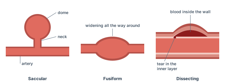
A dissecting aneurysm forms differently. The wall of an artery is made of several layers. A tear in the inner layer lets blood seep into the thickness of the wall, which it splits and makes bulge (Grand dictionnaire terminologique).
These aneurysms are rare in the brain. When they rupture, they often bleed again, sometimes within hours, which weighs heavily on the outcome; hence the value of very early treatment (Italian multicentre study, 2025). Because the wall of the artery itself is damaged, treatment often targets the artery, not just the pouch: in that study, about half of the endovascular treatments closed the diseased artery itself, the others used a stent that diverts the blood, or coils. Mine was treated with coils: see my story.
Causes and risk factors
Why the wall of an artery weakens is not fully understood. Some aneurysms come from a weakness present from birth. Several factors increase the risk:
- smoking;
- high blood pressure;
- excessive alcohol or drug use;
- a family history: when two or more first-degree relatives (parents, siblings, children) have had one, screening of the family is recommended;
- some inherited diseases of the connective tissue, such as Ehlers-Danlos syndromes or polycystic kidney disease;
- age, and being a woman.
More rarely, an aneurysm follows a head injury or an infection. The two factors you can act on are smoking and blood pressure.
Rupture: an emergency
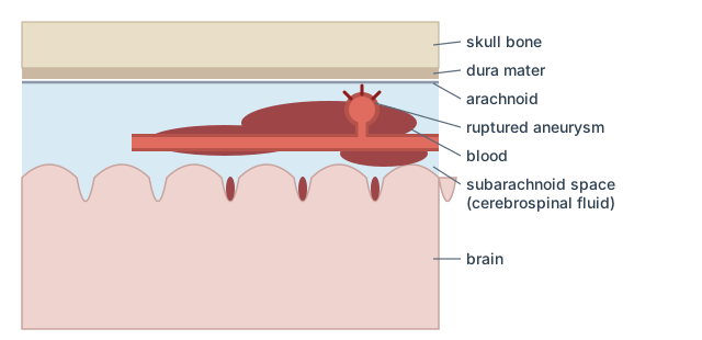
When an aneurysm ruptures, blood spreads between the brain and the membranes around it. This is a subarachnoid hemorrhage, a type of hemorrhagic stroke.
The signs appear suddenly:
- a violent headache that peaks within seconds, often described as a “thunderclap” or “the worst headache of my life”;
- nausea and vomiting;
- neck pain or stiffness;
- sensitivity to light, blurred vision;
- loss of consciousness, seizures.
Some people have an unusual “sentinel” headache hours or days before the rupture. Depending on the amount of blood, a rupture ranges from a mere stiff neck to coma, or even death. Frequent or chronic headaches, on the other hand, are not a sign of an aneurysm.
Diagnosis
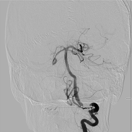
The first test is usually a CT scan, which shows the blood. A lumbar puncture can look for blood in the cerebrospinal fluid. To see the aneurysm itself, doctors use a CT angiography or an MR angiography, with an injection of contrast agent. The most precise test is cerebral angiography: a catheter inserted in the groin carries contrast agent directly into the arteries of the brain. A computer then “subtracts” the bones from the images, so that only the arteries remain.
Many unruptured aneurysms are found by chance, during a test done for another reason.
Treatment
The goal is to exclude the aneurysm from the blood flow so that it cannot bleed (again).
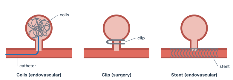
- Endovascular treatment (coils). A catheter inserted in the artery of the groin is guided up to the aneurysm, which is filled with fine coiled platinum wires. The blood clots inside and the aneurysm closes, without opening the skull. The coils stay in place for good. They can be placed during the same angiography that found the aneurysm.
- Stent. A small tube of metal mesh, placed by catheter, can divert the blood away from the aneurysm.
- Surgical clipping. The surgeon removes a piece of skull and places a metal clip at the base of the aneurysm.
A small unruptured aneurysm is often only monitored by imaging, while controlling the risk factors. The choice depends on the size, shape and location of the aneurysm, and on age and health. It is made with the neurosurgeon, by weighing the risk of treatment against the risk of rupture. After treatment, follow-up MRIs or angiographies check that the aneurysm stays closed.
After a rupture: complications
A ruptured aneurysm needs prompt care, because several complications can follow.
- Rebleeding. This is the main danger, especially in the first days. Without treatment, the risk reaches 40 to 50% within six months (CHUV).
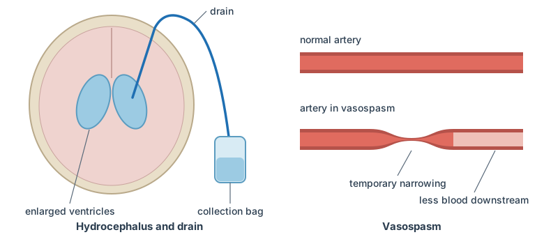
- Hydrocephalus. The blood can block the flow of cerebrospinal fluid, which builds up and compresses the brain. It then has to be drained, for example with a drain, as in my case.
- Vasospasm. In the following days, the arteries of the brain can narrow suddenly and temporarily. Part of the brain can then lack blood, which causes an ischemic stroke.
- Hyponatremia. The level of sodium in the blood can drop, which can make brain cells swell.
Recovery
The after-effects depend on the amount of blood and on the damage to the brain: cognitive and physical abilities, vision, balance, and more. Treating the aneurysm does not undo the effects of the hemorrhage. During recovery, some people have to learn new ways of doing everyday activities.
Sources and further reading
This page is based on these French-language resources, recommended by the Neuro Patient Resource Centre (consulted in November 2026):
- CHU de Québec – Université Laval, L’anévrisme cérébral non rompu
- Brain Injury Canada, Anévrismes cérébraux
- MSD Manual, Consumer Version, Anévrismes du cerveau
- CHUV, Lausanne University Hospital, L’anévrisme cérébral
- PasseportSanté, La rupture d’anévrisme
The section on dissecting aneurysms also relies on:
- The Neuro, InfoNeuro fact sheet Anévrisme cérébral (PDF, in French)
- Office québécois de la langue française and Royal College of Physicians and Surgeons of Canada, anévrisme disséquant, Grand dictionnaire terminologique
- Lozupone E. et al., Dissecting aneurysm in poor-grade subarachnoid hemorrhage, Journal of Neurosurgery, 2025
In English, the Centre also recommends:
- MSD Manual, Consumer Version, Brain Aneurysms
- Brain Injury Canada, Brain aneurysm
- The Brain Aneurysm Foundation, About Brain Aneurysms
- National Institute of Neurological Disorders and Stroke, Cerebral Aneurysms
- RadiologyInfo.org, Intracranial Vascular Treatments